


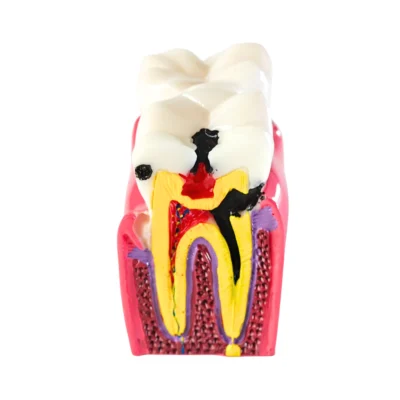
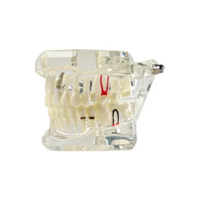
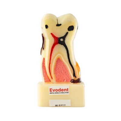
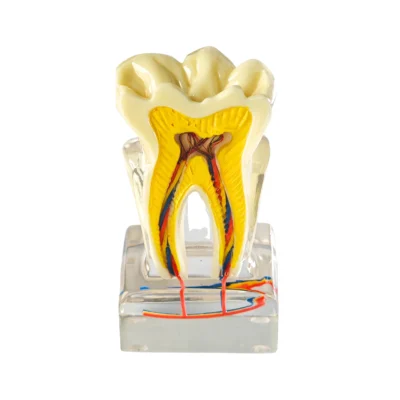
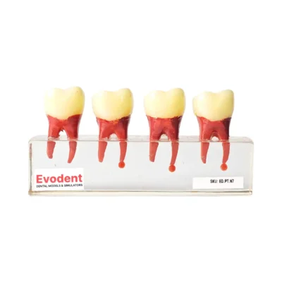
Evodent Advanced Tooth Decay Cross Section Model – Caries Progression, Pulp & Root Canal Anatomy
Original price was: ₹2,500.00.₹1,495.00Current price is: ₹1,495.00.
A detailed Tooth Decay Cross Section Model that clearly shows caries progression from enamel to pulp helping patients clearly understand pain, treatment and root canal necessity.
Shipping & Benefits
- Ships in 24 hours
- Damage-proof packaging
- GST invoice provided
Description
Explaining tooth decay to patients is harder than it should be.
Words like enamel, dentin, pulp and root canal often mean very little until patients can actually see what is happening inside the tooth. That’s exactly where the Evodent Advanced Tooth Decay Cross Section Model becomes invaluable.
This Evodent Advanced Tooth Decay Cross Section Patient Education Model presents a realistic cross section of a tooth, clearly showing how dental caries start on the surface and gradually progress deeper from enamel to dentin and finally into the pulp. Instead of abstract explanations, patients can visually follow the journey of decay, step by step. Dentists commonly use this model to reduce chairside explanation time and improve treatment acceptance during cavity and RCT discussions.
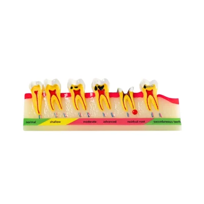
What This Tooth Decay Cross Section Model Shows
This model is designed to make complex dental concepts instantly understandable:
- The outer enamel layer and where cavities usually begin
- The dentin layer explaining sensitivity and rapid decay spread
- The pulp chamber showing why pain and infection occur
- Advanced caries progression including deep lesions reaching the nerve
- Root canal anatomy helping patients understand endodontic treatment
The use of high-contrast colors makes the difference between healthy tissue and decayed areas immediately obvious even to children or anxious patients.
Why This Model Is Excellent for Patient Education
Most patients struggle to visualize what is happening inside their tooth. X-rays can be confusing, and verbal explanations often fall flat. This model helps by:
- Turning invisible problems into visible ones
- Reducing patient confusion, fear and treatment hesitation
- Improving treatment acceptance for fillings, RCT and crowns
- Making explanations faster and more effective
- Helping patients understand urgency and consequences
When patients see decay reaching the pulp, they immediately understand why procedures like fillings, root canals or crowns are necessary.
How This Model Is Commonly Used in Clinics
- Chairside explanation during consultations
- Educating patients about cavities and decay progression
- Explaining pain, sensitivity and infection causes
- Demonstrating why early treatment matters
- Explaining why a root canal is required
- Educating children, first-time patients and nervous patients
Build Quality & Visual Accuracy
- Durable solid construction for repeated daily use
- Clear, bold color coding for enamel, dentin, pulp and decay
- Smooth, professional finish suitable for clinic desks
- Designed for long term use
The model is built to withstand daily patient demonstrations without wear or fading.
Technical Specifications
- Product Name: Evodent Advanced Tooth Decay Cross Section Model
- Type: Tooth Decay / Caries Cross Section Model / Root Canal Model
- Material: High quality durable resin
- Design: Cross section showing internal tooth anatomy
- Anatomy Shown: Enamel, dentin, pulp chamber, root canal
- Caries Representation: Early to advanced decay stages
- Finish: Hand-painted, high-contrast anatomical colouring
- Scale: Enlarged for clear visibility
- Color Coding: High contrast anatomical colors
- Primary Use: Patient education and chairside demonstration
- Application: Dental clinics, counselling, awareness programs
- Placement: Consultation desk or chairside
- Maintenance: Easy to clean with a soft damp cloth
Ideal For
- Dental clinics and private practices
- Patient education and counselling
- Explaining caries, cavities and tooth pain
- Root canal and restorative treatment explanations
- Chairside demonstrations
- Dental awareness programs
FAQs – Tooth Decay Cross Section Model
Q. What is this tooth cross section model used for?
A. It is used to visually explain tooth decay, cavities, internal tooth anatomy and caries progression to patients in a simple and clear way.
Q. Does it show real stages of tooth decay?
A. Yes. It clearly shows how decay progresses from enamel to dentin and finally to the pulp.
Q. Can this model help explain tooth pain to patients?
A. Yes, it visually explains why pain occurs when decay reaches deeper layers or the nerve.
Q. Is this useful for explaining root canal treatment?
A. Yes. The exposed pulp makes it very effective for explaining why root canal treatment is required.
Q. Is this model suitable for children and nervous patients?
A. Yes. The clear colors and simple design make it easy to understand without being intimidating.
You may also like…
-
Sale!
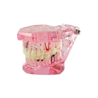

Evodent All in One Model, Pink
Original price was: ₹5,000.00.₹1,195.00Current price is: ₹1,195.00. -
Sale!

Evodent Signature All in One with Three Implant
Original price was: ₹5,000.00.₹1,990.00Current price is: ₹1,990.00. -
Sale!

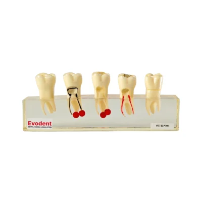
Evodent Tooth Cross-Section Model – Caries, Pulpitis, Periodontal Infection and Lesion Progression (Patient Education Model)
Original price was: ₹3,500.00.₹1,800.00Current price is: ₹1,800.00. -
Sale!

Evodent Tooth Anatomy Cross-Section Model with Pulp and Nerve Supply for Patient Education
Original price was: ₹3,000.00.₹1,995.00Current price is: ₹1,995.00.
Related products
-
Sale!
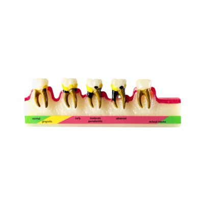

Evodent Periodontal Disease Progression Model – Gingivitis to Advanced Periodontitis & Occlusal Trauma
Original price was: ₹6,000.00.₹1,995.00Current price is: ₹1,995.00. -
Sale!

Evodent Root Canal Treatment RCT Stages Model – Caries, Pulp Exposure, Endodontic Filling and Post Core Demonstration
Original price was: ₹6,000.00.₹1,500.00Current price is: ₹1,500.00. -
Sale!

Evodent Signature Tooth Decay & Caries Progression Model – 6 Stage Patient Education Model
Original price was: ₹6,000.00.₹1,995.00Current price is: ₹1,995.00. -
Sale!

Evodent Root Canal Treatment Stages Model – Caries Progression, Pulp Infection, Endodontic Instrumentation and Gutta-Percha Filling Demonstration
Original price was: ₹6,000.00.₹1,500.00Current price is: ₹1,500.00.