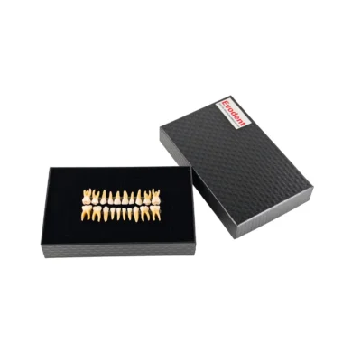
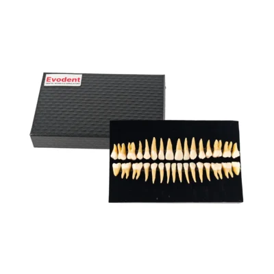
Evodent Tooth Cross Section Model – Caries, Pulpitis, Periodontal Infection and Lesion Progression for Teaching, for Dental Education & Patient Explanation
$39.99 USD
A tooth cross section model used to demonstrate dental anatomy, caries progression, and root canal structure for teaching and patient education.
Shipping & Benefits
- International shipping available
- Tracking details shared by email
- Trusted by dental professionals worldwide
Description
Explaining what happens inside a tooth is difficult without a visual reference. Concepts like caries progression, pulp involvement and root canal pathways are not easy to communicate using only diagrams or verbal explanation.
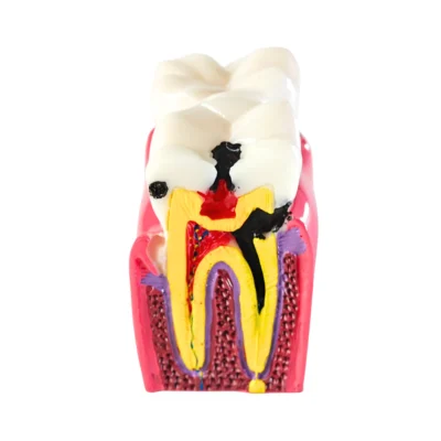
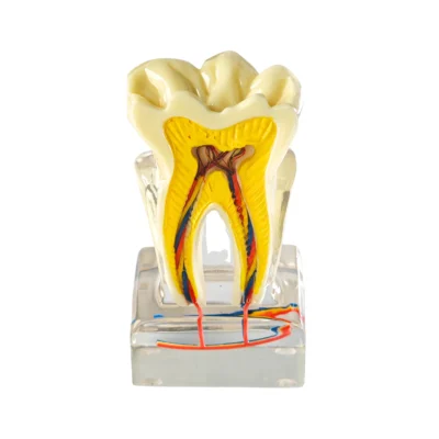
This tooth cross section model is designed to make internal tooth anatomy clearly visible. It shows how decay progresses, how the pulp is affected and how root canal structures are arranged all in a single physical model.
This makes it useful in both teaching environments and clinical explanation settings, where clarity and visual understanding are important.
What is a tooth cross section model?
A tooth cross section model is a dental anatomy model that shows the internal structure of a tooth in a cut section view, including enamel, dentin, pulp, and root canal pathways. It is commonly used to explain caries progression, pulpitis, and root canal treatment in both teaching and clinical settings. This includes:
- Enamel and dentin layers
- Pulp chamber and root canal pathways
- Caries (decay) progression
- Surrounding supporting structures
This Evodent model is designed as a static educational model, used for demonstration and explanation rather than procedural training.
This type of model is also referred to as a:
- Dental anatomy model
- Tooth anatomy teaching model
- Caries progression model
- Root canal anatomy demonstration model
These terms are often used interchangeably depending on whether the focus is structure, pathology, or clinical explanation.
What does this tooth cross section model demonstrate clinically?
This model visually represents the internal structure of a tooth along with disease progression stages that are directly relevant in clinical practice:
- Enamel, dentin, and pulp anatomy in cross-section
- Caries progression from surface to pulp involvement
- Root canal pathway extending through the root
- Pulp infection and lesion spread
- Surrounding bone and support structures
This allows simultaneous understanding of structure + pathology, which is difficult to achieve using flat diagrams.
How does caries progress inside a tooth? (Visual explanation using this model)
Caries begins at the enamel surface and gradually penetrates into dentin. If untreated, it reaches the pulp, causing inflammation (pulpitis) and eventually infection.
This model helps demonstrate:
- Early enamel decay
- Dentin involvement
- Pulp exposure and infection
- Need for root canal treatment
This makes it especially useful for explaining why treatment is required, not just what the problem is.
Specialty Relevance
This model is relevant across multiple dental domains:
- Endodontics: Understanding pulp chamber and root canal anatomy
- General Dentistry: Explaining caries progression and treatment planning
- Pedodontics: Teaching basic tooth structure and decay concepts
- Patient Education: Demonstrating why treatments like root canal are required
How is this model used in dental clinics and colleges?
In teaching environments:
- Teaching internal tooth anatomy in 3D
- Explaining caries progression step-by-step
- Demonstrating pulp and root canal structures
In clinics:
- Explaining root canal treatment to patients
- Showing how decay spreads internally
- Improving case acceptance through visual clarity
Who is this for?
- Dental students learning anatomy and pathology
- Dentists explaining treatment plans to patients
- Training institutes and skill labs
- Clinics that use visual tools for patient communication
What makes this model effective for patient communication?
Patients often struggle to understand internal tooth problems because they cannot see them. This model solves that by:
- Making invisible structures visible
- Showing depth of decay clearly
- Connecting symptoms to internal damage
- Helping patients understand treatment necessity
This improves communication and can help patients make more informed decisions.
Why use a physical cross section model instead of diagrams?
- Provides 3D understanding of internal structures
- Makes it easier to explain depth and progression of decay
- Helps patients and students grasp concepts faster
- Allows repeated use in both teaching and clinical settings
Technical Specifications
- Model Type: Tooth cross section model
- Category: Dental anatomy / pathology demonstration model
- Structure Shown:
- Enamel
- Dentin
- Pulp chamber
- Root canal pathway
- Pathology Representation:
- Caries progression
- Pulp involvement
- Mounting: Fixed base (non-removable tooth)
- Usage Type: Demonstration and explanation
- Interaction: Visual (no operative use)
Tooth Cross Section Model vs Typodont vs Dental Simulator
A tooth cross section model is different from typodonts and dental simulators in how it is used.
- Tooth cross section model: Used for visual explanation of internal tooth anatomy and disease progression
- Typodont: Used for hands-on practice like cavity preparation, restorations, or orthodontics
- Dental simulator (phantom head): Used for full clinical simulation with instruments
This Evodent model is a demonstration model, not a procedural training tool.
How accurate is this model compared to real tooth anatomy?
This model is designed to represent clinically relevant anatomical structures and disease progression, not exact microscopic detail. It simplifies complex anatomy into a clear visual format, making it easier to teach and explain key concepts.
How this model is typically used?
Placed on a desk or chairside during explanation Used alongside verbal explanation of:
- Tooth structure
- Decay progression
- Root canal treatment steps
- Referenced repeatedly during teaching or consultation
Maintenance
- Can be cleaned with a dry or slightly damp cloth
- No special storage requirements
FAQs – Evodent Tooth Cross Section Model
Q. What is a tooth cross section model used for in dentistry?
A. A tooth cross section model is used to demonstrate internal tooth anatomy and disease progression such as caries and pulp involvement.
Q. How does this model help in patient education?
A. It helps patients visually understand how decay progresses and why treatments like root canal are required, improving communication and decision-making.
Q. Does this model show root canal anatomy?
A. Yes, the model clearly shows the root canal pathway and pulp chamber, making it useful for explaining endodontic concepts.
Q. Is this model used for practicing dental procedures?
A. No, this is a demonstration model only and is not designed for drilling, instrumentation, or procedural training.
Q. What is the difference between a tooth model and a typodont?
A. A tooth model is used for visual explanation and teaching, while a typodont is used for hands-on procedural training.
Q. Does this model show caries progression clearly?
A. Yes, the model visually represents how decay progresses from enamel to dentin and pulp, making it useful for both teaching and patient explanation.
Q. Is this a root canal training model?
A. No, this is a demonstration model used to explain root canal anatomy, not for performing procedures.
Q. What type of tooth does this model represent?
A. The model represents a molar tooth with internal anatomy and multi-layer structure.
Q. Which subjects or specialties is this model useful for?
A. It is useful in endodontics, general dentistry, pedodontics, and patient education.
Q. Can this model be used in clinics?
A. Yes, it is commonly used in clinics for explaining treatment plans and dental conditions to patients.
Why this model matters in dental education and practice
Understanding internal tooth anatomy is fundamental to diagnosing and explaining dental conditions. A physical cross section model bridges the gap between theory and real-world understanding by making complex structures easy to visualize.