Evodent Dental Caries Progression Model – Tooth Decay & Patient Education Model
Original price was: ₹6,000.00 INR.₹1,799.00 INRCurrent price is: ₹1,799.00 INR.
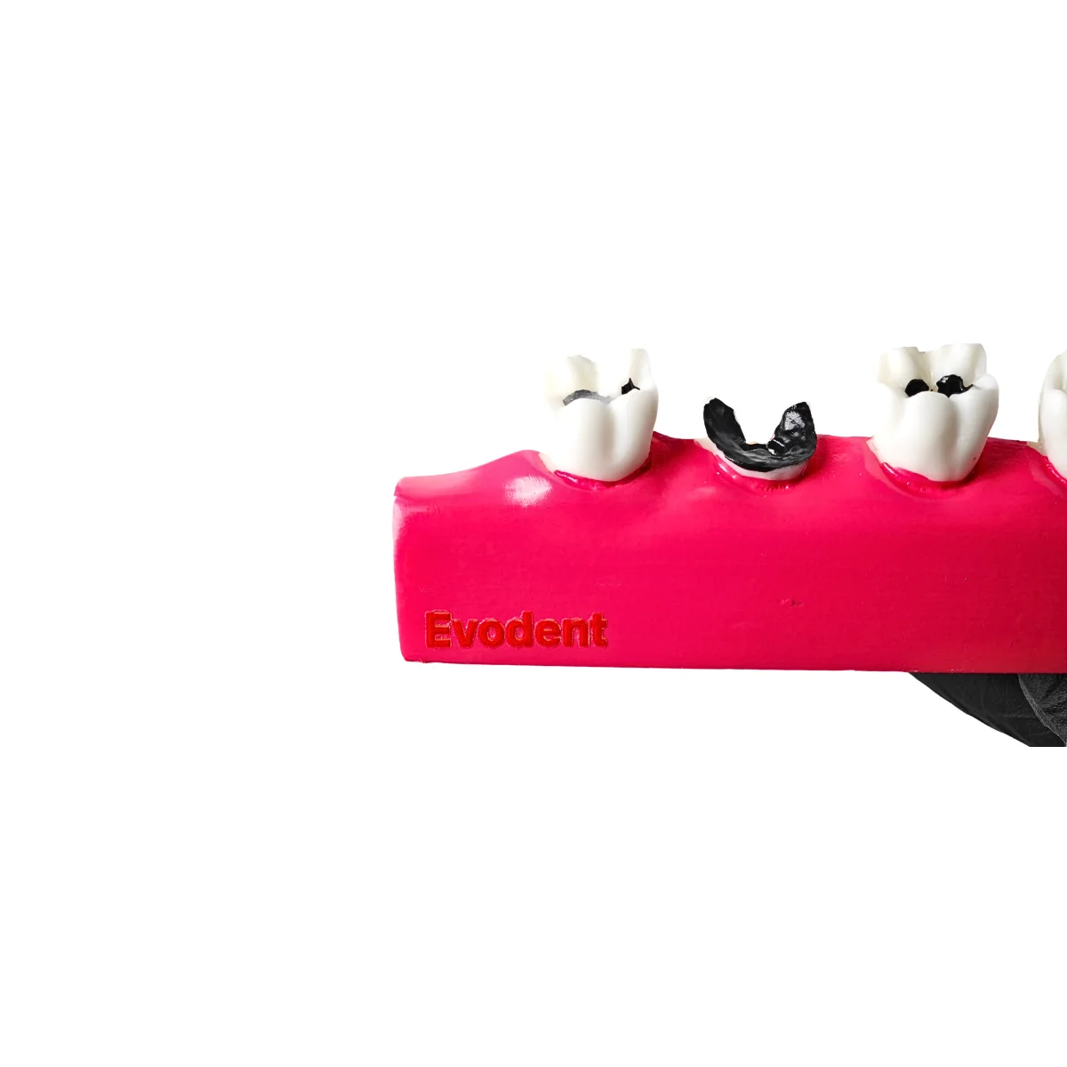
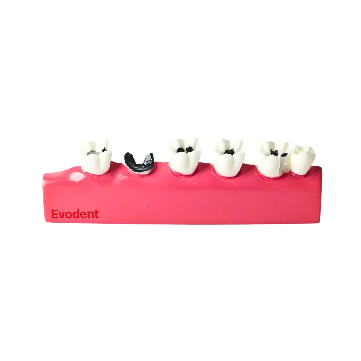
A cross-section dental caries model showing healthy tooth structure, progressive decay, residual root and a succedaneous-tooth example for clear patient education and dental teaching.
Shipping & Benefits
- International shipping available
- Tracking details shared by email
- Trusted by dental professionals worldwide
Description
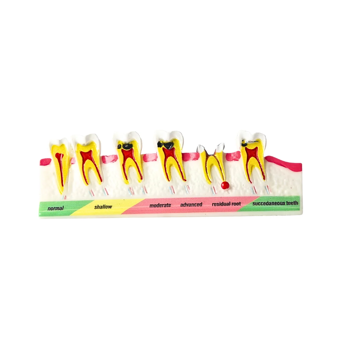
The Evodent Dental Caries Progression Model is a cross-section dental model designed to make tooth decay easier for patients and students to understand.
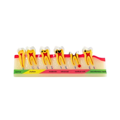
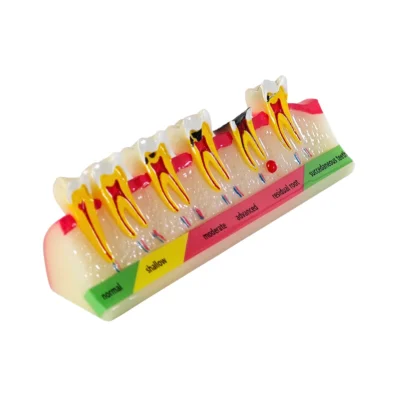
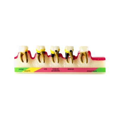
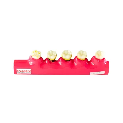
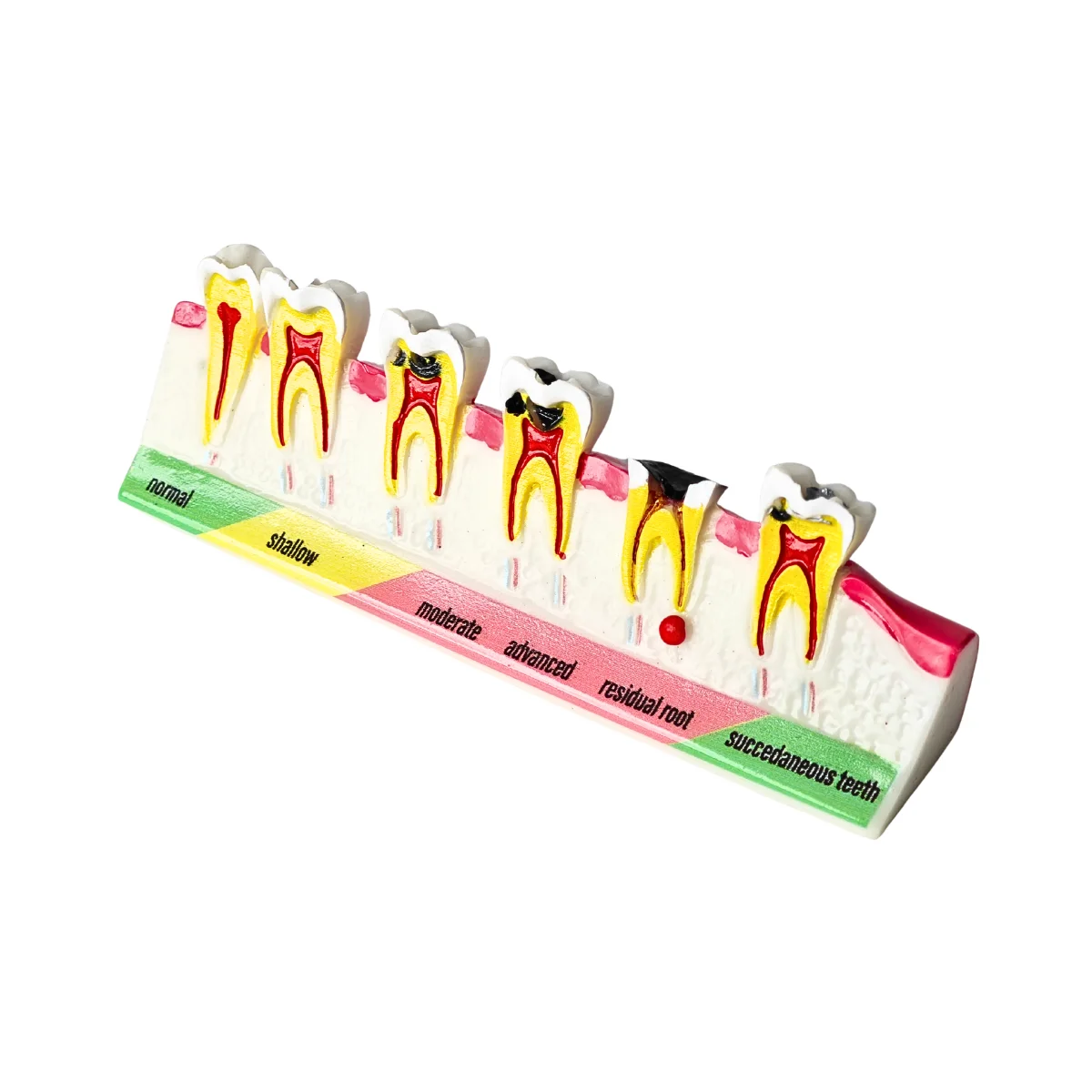
Rather than showing only the outside of the tooth, the model exposes the internal tooth structure and places different conditions side by side. This allows the dentist or educator to visually move from a healthy tooth through progressively deeper caries and then discuss more extensive tooth destruction, a residual root and a succedaneous-tooth example.
The labelled, color-coded base makes it easy to follow during a chairside explanation.
It is designed primarily for patient education, with additional value for dental students, oral-health education and classroom demonstrations.
Explain Tooth Decay in a Way Patients Can See
Patients often understand the word cavity but may not understand what happens as dental caries moves deeper into a tooth. A dental caries progression model gives the dentist a three-dimensional reference while explaining the condition. It can help demonstrate:
- The appearance of a healthy tooth
- Early or shallow carious involvement
- Progressively deeper tooth decay
- The relationship between the cavity and internal tooth structures
- Advanced destruction of tooth structure
- What may remain when much of the crown has been lost
- A residual root condition
- The relationship between primary teeth and a succedaneous tooth in the illustrated example
Because the examples are positioned next to one another, patients can compare them immediately instead of trying to understand several separate diagrams.
What Does the Dental Caries Model Show?
The model uses labelled sections to demonstrate different dental conditions.
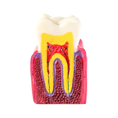
- Normal Tooth: The normal tooth provides the reference point. It allows the dentist to first show healthy tooth structure before moving through the examples of disease.
- Shallow Caries: The model then introduces a relatively shallow carious lesion. This is useful when explaining that decay can begin at the tooth surface and may be much less extensive in its earlier form.
- Moderate Caries: The moderate example shows a deeper lesion and makes the progression of the disease easier to visualize. Patients can directly compare it with the shallow example beside it.
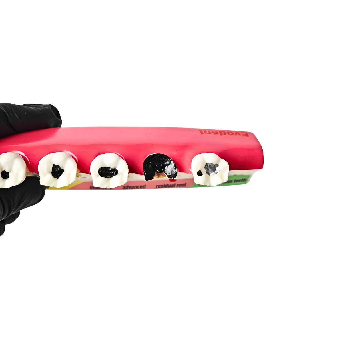
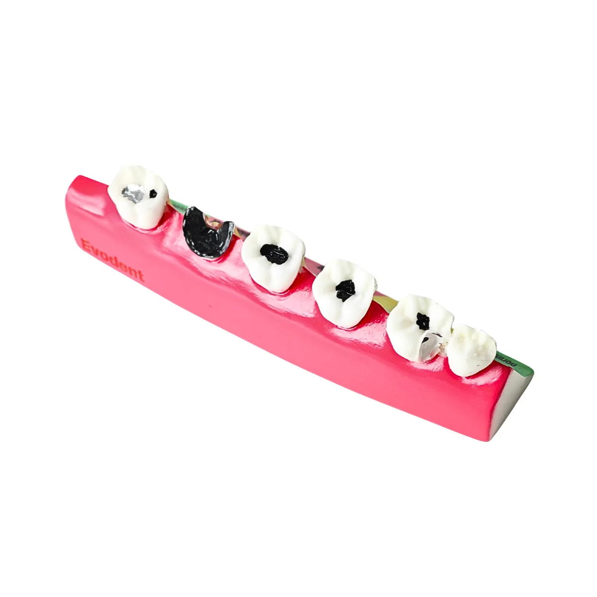
- Advanced Caries: The advanced section shows more extensive destruction of the tooth. The cross-section format helps demonstrate that an apparently visible cavity may involve deeper internal structures than the patient can see from the outside.
- Residual Root: The residual-root example shows a condition in which much of the coronal tooth structure has been lost and a root portion remains. This is particularly useful when explaining why a severely broken-down tooth may be very different from a small cavity.
- Succedaneous Tooth Example: The final section includes a succedaneous-tooth example. This should be explained as a separate developmental/dental condition shown on the model, rather than as the next stage of dental caries.
Cross-Section Design Shows What Happens Inside the Tooth
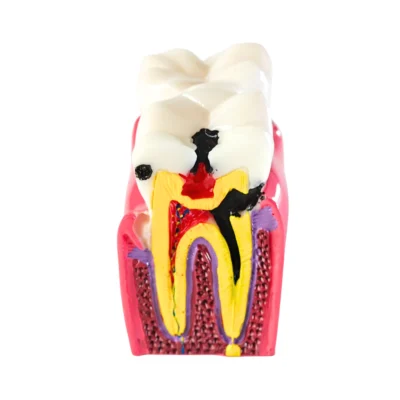
One of the most useful features of this model is the cutaway design. Patients usually see only the crown of their own tooth. The cross-section gives the dentist a way to point to structures inside and below the crown while explaining what is happening.
The model visibly represents:
- Tooth crown
- Root structure
- Internal pulp space
- Surrounding supporting structure
- Increasing areas of carious destruction
- Residual root condition
This makes it useful when discussing why treatment needs can change depending on how deeply a tooth is affected.
A dentist can use the model alongside the patient’s own radiographs, photographs or examination findings rather than asking the patient to understand the problem from an X-ray alone. Comparable professional patient-education caries models are specifically intended to be used alongside patient radiographs and images for this purpose.
Useful for Patient Education
Patient communication is the main reason to use this model. It can support conversations about:
- Dental caries and cavities
- Early versus more extensive decay
- Why early diagnosis matters
- Why two cavities may require different treatment
- Progression of untreated tooth decay
- Loss of tooth structure
- Residual roots
- Oral hygiene and prevention
- The need for regular dental examinations
- Treatment discussions based on the patient’s actual diagnosis
A simple approach is to start at the healthy tooth and move across the model while explaining how the condition becomes more extensive. This gives the patient a visual story rather than a list of technical terms.
How to Use the Model During a Patient Consultation
You do not need a long explanation.
- Start with the normal tooth to establish what healthy tooth structure looks like.
- Move through shallow, moderate and advanced caries while pointing out how the affected area becomes more extensive.
- Use the cross-section to explain that decay can affect structures patients cannot see from the outside.
- Show the residual root example when discussing severely broken-down teeth or retained roots.
- Relate the model back to the patient’s own findings using their examination, radiographs or intraoral photographs.
The model is a general educational aid. It should support, not replace, the dentist’s diagnosis and patient-specific explanation.
Useful for Dental Students & Teaching
Although designed primarily for patient education, the model is also useful for:
- Dental students
- Dental assistants
- Dental hygienists
- Dental colleges
- Oral-health educators
- School dental-health programs
- Classroom demonstrations
- Dental training institutes
Students can use it to compare visible caries progression and understand the relationship between external tooth destruction and internal anatomy. It is a visual demonstration model, not a procedural cavity-preparation or restorative practice model.
Key Features
- Cross-section dental caries progression model
- Healthy / normal tooth reference
- Shallow caries representation
- Moderate caries representation
- Advanced caries representation
- Residual root example
- Succedaneous-tooth example
- Internal tooth structures visible
- Carious areas highlighted for easier explanation
- Labelled, color-coded condition guide
- Designed primarily for patient education
- Useful for dental teaching and oral-health education
- Side-by-side design for easy comparison
- Freestanding model for chairside or classroom use
- Reusable for repeated patient consultations
Technical Specifications
- Product Type: Dental caries patient education model
- Model Type: Dental caries / tooth decay progression model
- Design: Cross-sectional dental demonstration model
- Conditions Shown: Normal, shallow caries, moderate caries, advanced caries, residual root and succedaneous-tooth example
- Internal Tooth Anatomy: Visible
- Condition Identification: Labelled and color-coded base
- Primary Use: Patient education
- Secondary Use: Dental teaching and oral-health education
- Suitable For: Dentists, dental students, hygienists, educators, clinics and training institutes
- Use Environment: Chairside consultation, dental clinic, classroom or training room
- Practice Type: Visual education and demonstration; not intended for procedural cavity preparation
FAQs – Evodent Dental Caries Progression Model
Q. What is a dental caries progression model used for?
A. A dental caries progression model is used to visually explain how tooth decay can become more extensive. This model is primarily intended for patient education and allows dentists to compare a healthy tooth with different examples of carious destruction.
Q. What conditions are shown on this tooth decay model?
A. The model shows a normal tooth, shallow caries, moderate caries, advanced caries, a residual root and a succedaneous-tooth example.
Q. Does this model show the inside of the tooth?
A. Yes. The cross-section design shows the internal tooth and root structures, making it easier to explain how dental caries can extend beyond what is visible on the outer surface.
Q. Is this model suitable for explaining cavities to patients?
A. Yes. Patient education is its primary use. Dentists can move through the labelled examples while explaining how a small or shallow lesion differs from more extensive tooth decay.
Q. What is a residual root on the model?
A. The residual-root section demonstrates a condition where much of the visible crown has been lost while a root portion remains. It can be used when explaining severely broken-down teeth or retained roots.
Q. Is the succedaneous tooth shown as a stage of tooth decay?
A. No. The succedaneous-tooth example is shown separately on the model and should not be described as the final stage of caries progression.
Q. Can this dental caries model be used with patient X-rays?
A. Yes. It can be used as a general visual reference while discussing a patient’s radiographs, photographs or examination findings. The model helps explain the concept, while the patient’s actual diagnosis should be based on their clinical findings.
Q. Can dental students use this model?
A. Yes. It is also useful for dental students, hygienists and educators studying or teaching tooth anatomy, caries progression and patient communication.
You may also like…
- Sale!

Evodent Large Crystal Dental Implant Demonstration Model – Removable Crown and Implant Components
Original price was: ₹3,000.00 INR.₹1,995.00 INRCurrent price is: ₹1,995.00 INR. - Sale!
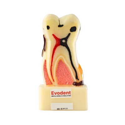
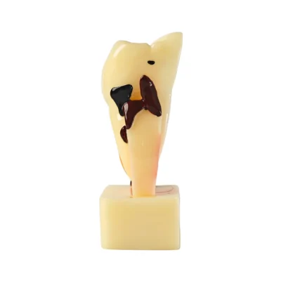
Evodent Tooth Cross Section Model – Caries, Pulpitis, Periodontal Infection and Lesion Progression for Teaching, for Dental Education & Patient Explanation
Original price was: ₹3,500.00 INR.₹1,800.00 INRCurrent price is: ₹1,800.00 INR. - Sale!
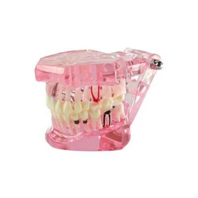
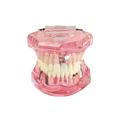
Evodent All in One Model, Pink – Implants, Crown, Caries, Bridges, Impacted Teeth and Full Treatment Demonstration Model
Original price was: ₹5,000.00 INR.₹1,295.00 INRCurrent price is: ₹1,295.00 INR. - Sale!
Evodent Dental Typodont Model with Soft Gingiva and Removable Teeth, Dental Extraction Demonstration Model with Root Anatomy for Patient Education
Original price was: ₹5,000.00 INR.₹2,995.00 INRCurrent price is: ₹2,995.00 INR.
Related products
- Sale!
Evodent Periodontal Disease Progression Model – Gingivitis, Periodontitis & Occlusal Trauma
Original price was: ₹6,000.00 INR.₹1,799.00 INRCurrent price is: ₹1,799.00 INR. - Sale!
Evodent Signature Tooth Decay & Caries Progression Model – 6 Stage Patient Education Model
Original price was: ₹6,000.00 INR.₹1,995.00 INRCurrent price is: ₹1,995.00 INR. - Sale!
Evodent Periodontal Disease Progression Model – Gingivitis to Advanced Periodontitis & Occlusal Trauma
Original price was: ₹6,000.00 INR.₹1,995.00 INRCurrent price is: ₹1,995.00 INR. - Sale!
Evodent Advanced Tooth Decay Cross Section Model – Caries Progression, Pulp & Root Canal Anatomy
Original price was: ₹2,500.00 INR.₹1,495.00 INRCurrent price is: ₹1,495.00 INR.