


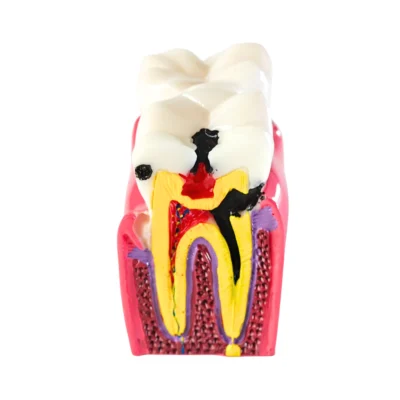
Evodent Tooth Anatomy Cross-Section Model with Pulp and Nerve Supply for Patient Education
Original price was: ₹3,000.00.₹1,995.00Current price is: ₹1,995.00.
Tooth Anatomy Cross-Section Model designed to clearly demonstrate internal tooth structure, pulp chamber and nerve supply for patient education.
Shipping & Benefits
- Ships in 24 hours
- Damage-proof packaging
- GST invoice provided
Description
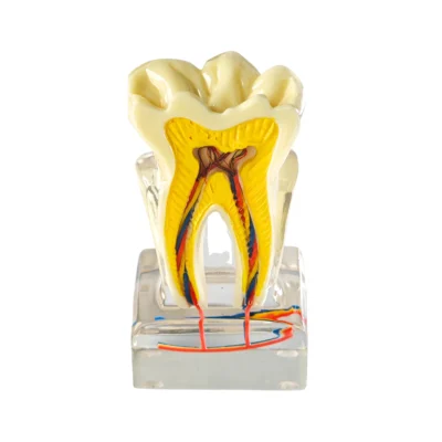
The Tooth Anatomy Cross-Section Model is a visual demonstration model designed to clearly explain the internal structure of a human tooth in a way that is easy to understand, remember and explain. By showing a detailed cross section of the tooth, this model helps bridge the gap between textbook diagrams and real-world dental conversations.
Unlike external tooth models that only show crown shape, this tooth anatomy cross-section model reveals what’s happening inside the tooth including enamel, dentin, pulp chamber, root canals and nerve supply. This makes it especially useful when explaining tooth pain, sensitivity, cavities, root canal treatment or basic dental anatomy to patients and students.
The internal anatomy is color-coded and clearly separated, allowing viewers to immediately identify each layer of the tooth. The pulp chamber and nerve pathways are visibly highlighted, making it easier to explain why certain dental conditions cause pain and why procedures like root canal treatment are necessary.
Because the model is mounted on a stable transparent base, it can be comfortably used during chairside consultations, classroom teaching or patient education sessions without needing additional diagrams or screens.
Why patients struggle to understand tooth pain (and how this model helps)?
Most patients experience tooth pain without understanding where it comes from. Terms like “pulp,” “nerve,” or “root canal” sound abstract when explained verbally.
The Tooth Anatomy Cross-Section Model solves this by visually showing exactly where pain originates, how infection spreads, and why certain treatments are recommended. When patients see the pulp chamber and nerve pathways, explanations become intuitive instead of overwhelming.
What does this Tooth Anatomy Cross-Section Model show?
This Tooth Anatomy Cross-Section Model visually demonstrates:
- Enamel (outer protective layer of the tooth)
- Dentin (supporting layer beneath enamel)
- Pulp chamber with nerve and blood supply
- Root canal anatomy extending into the roots
- Internal tooth morphology in a single cross-sectional view
This makes complex dental anatomy simple to explain even to patients with no medical background.
Why this model is useful for patient education
Many patients struggle to understand dental problems when explanations are purely verbal. Showing a tooth anatomy cross-section model instantly improves understanding by allowing patients to see where pain, infection, or damage occurs.
This model helps:
- Explain tooth pain and sensitivity clearly
- Demonstrate why cavities progress deeper over time
- Visually support root canal treatment discussions
- Improve patient confidence and treatment acceptance
When patients understand what’s happening inside the tooth, conversations become easier and more productive.
Who is this Tooth Anatomy Cross-Section Model for?
This model is ideal for:
- Dental clinics for chairside patient explanation
- Dentists and endodontists explaining pulp and nerve-related issues
- Dental colleges and training institutes for anatomy teaching
- Patient education areas in dental clinics or dental hospitals
How this model is commonly used
The Tooth Anatomy Cross-Section Model is commonly used to:
- Explain internal tooth anatomy during consultations
- Show patients where nerve pain originates
- Support discussions about root canal treatment
- Teach basic tooth structure in classrooms
- Act as a physical reference during dental education sessions
It serves as a simple, reliable visual aid that reduces confusion and improves communication.
Key Features at a Glance
- Clear tooth anatomy cross-section design
- Visible enamel, dentin, pulp wand nerve supply
- Color-coded internal structures for easy explanation
- Stable transparent base for repeated use
- Designed specifically for dental education and patient communication
Technical Specifications
- Product Name: Tooth Anatomy Cross-Section Model
- Product Type: Dental anatomy demonstration model
- Model Design: Cross-sectional tooth anatomy display
- Anatomy Shown: Enamel, dentin, pulp chamber, nerve supply, root canals
- Purpose: Patient education and dental teaching
- Use Environment: Dental clinics, dental colleges, teaching labs
- Model Type: Fixed, non-removable anatomy model
- Base Type: Transparent acrylic base
- Color Coding: Yes (for internal anatomy identification)
- Intended Use: Visual explanation of tooth structure and nerve anatomy
- Clinical Use: Not intended for clinical or procedural use
This model is designed based on standard dental anatomy principles commonly taught in dental education and used in daily clinical communication, making it a reliable and practical reference tool for dental professionals.
FAQs – Tooth Anatomy Cross-Section Model
Q. What is a Tooth Anatomy Cross-Section Model used for?
A. The Tooth Anatomy Cross-Section Model is used to visually explain the internal structure of a tooth, including enamel, dentin, pulp chamber, root canals and nerve supply, during patient education and dental teaching.
Q. Is this Tooth Anatomy Cross-Section Model suitable for patient education?
A. Yes, the model is specifically designed for patient education, helping patients understand tooth pain, sensitivity, cavities and root canal treatment through clear visual explanation.
Q. Can this model be used to explain root canal treatment?
A. Yes, the Tooth Anatomy Cross-Section Model helps explain where the pulp and nerves are located and why root canal treatment may be required when the pulp becomes infected or damaged.
Q. Who commonly uses a Tooth Anatomy Cross-Section Model?
A. This model is commonly used by dentists, endodontists, dental clinics, dental colleges, and teaching institutes for explaining internal tooth anatomy.
Q. Does this model show nerve supply inside the tooth?
A. Yes, the model clearly highlights the nerve supply within the pulp chamber and root canals, making it easier to explain tooth pain and sensitivity.
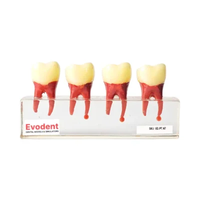
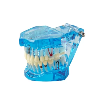
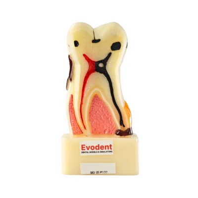
You may also like…
-
Sale!

Evodent Tooth Cross-Section Model – Caries, Pulpitis, Periodontal Infection and Lesion Progression (Patient Education Model)
Original price was: ₹3,500.00.₹1,800.00Current price is: ₹1,800.00. -
Sale!

Evodent All in One Model, Blue
Original price was: ₹5,000.00.₹1,195.00Current price is: ₹1,195.00. -
Sale!
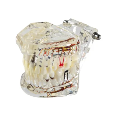

Evodent All in One Dental Implant and Nerve Anatomy Patient Education Model – Transparent Jaw, Teeth, Roots & Nerve Supply
Original price was: ₹5,000.00.₹1,495.00Current price is: ₹1,495.00. -
Sale!

Evodent Advanced Tooth Decay Cross Section Model – Caries Progression, Pulp & Root Canal Anatomy
Original price was: ₹2,500.00.₹1,495.00Current price is: ₹1,495.00.