


Evodent Orthognathic and Osteology Model for TMJ Anatomy, Jaw Movement and Maxillofacial Relationship Demonstration
Original price was: ₹25,000.00 INR.₹19,990.00 INRCurrent price is: ₹19,990.00 INR.
Interactive orthognathic anatomy model with TMJ movement, implant, sinus, nerve supply and maxillofacial relationships.
Shipping & Benefits
- International shipping available
- Tracking details shared by email
- Trusted by dental professionals worldwide
Description
Some treatment concepts are too complex to explain with CBCT scans, 2D images or static skull models alone.
The Evodent Orthognathic and Osteology Model is a premium surgical communication and patient education system designed for clinicians who need to visually explain TMJ anatomy, jaw movement, Class I, Class II, and Class III skeletal relationships, orthognathic surgery, implant positioning, sinus lift procedures, nerve anatomy and maxillofacial treatment planning with confidence.
Built for advanced chairside communication, this model combines dynamic mandibular movement, a removable magnetic maxilla, visible implant positioning, sinus anatomy, nerve pathways, nasal cavity anatomy, and skeletal landmark visualization in one interactive anatomical system.
Unlike static skull models, this interactive orthognathic model combines:
- Fully movable mandible
- Magnetic removable maxilla
- Class I, II and III skeletal relationship demonstration
- TMJ articulation
- Implant positioning visualization
- Maxillary sinus anatomy
- Inferior alveolar nerve pathways
- Visible nasal cavity anatomy
This allows clinicians to demonstrate not just describe how anatomy directly influences treatment decisions.
What can this orthognathic model demonstrate?
This premium maxillofacial model allows clinicians to visually demonstrate:
Skeletal relationships
- Class I occlusion
- Class II malocclusion
- Class III malocclusion
- Maxilla–mandible discrepancies
- Bite relationship changes
TMJ and jaw movement
- Mandibular opening and closing
- Functional jaw movement
- TMJ articulation
- Occlusal repositioning
Implant and sinus planning
- Dental implant positioning
- Implant-to-bone relationship
- Maxillary sinus anatomy
- Sinus lift planning concepts
- Surgical safety zones
Surgical communication
- Orthognathic treatment concepts
- Maxillary repositioning
- Mandibular repositioning
- Skeletal correction planning
- Post-surgical occlusal relationships
Internal anatomy
- Inferior alveolar nerve pathway
- Nasal cavity anatomy
- Maxillary sinus visualization
- Adjacent craniofacial bone relationships
What patients can physically see and understand
With this model, patients can clearly visualize:
- Why the upper and lower jaws do not align
- How Class II and Class III bite discrepancies occur
- Where implants are positioned
- How close implants are to the sinus floor
- Why sinus lift procedures may be necessary
- How jaw repositioning changes facial balance
- here major nerve pathways must be protected
When patients see anatomy, treatment conversations become easier.
Key interactive features
Movable mandible
The mandible is fully movable, allowing clinicians to demonstrate:
- Jaw opening and closing
- Functional occlusion
- TMJ movement
- Bite relationship changes
- Pre- and post-surgical jaw positioning concepts
Magnetic removable maxilla
The maxilla can be removed and repositioned using magnetic fixation, making it easier to explain:
- Orthognathic treatment planning
- Skeletal discrepancies
- Surgical repositioning
- Anatomical access to internal structures
Visible internal anatomy
Internal anatomical landmarks are clearly visible, including:
- Maxillary sinus
- Nasal cavity
- Nerve pathways
- Implant placement zone
- Adjacent skeletal anatomy
Who is this model for?
This Evodent Orthognathic and Osteology Model is designed for:
- Oral and maxillofacial surgeons
- Implantologists
- Prosthodontists
- Orthodontists
- Dental educators
- Dental colleges
- Multi-specialty clinics
- Surgical consultation centers
- Advanced implant education programs
Especially useful for:
- Orthognathic surgery consultations
- Class II / Class III case acceptance
- Implant treatment discussions
- Sinus lift planning discussions
- TMJ patient communication
- Skeletal discrepancy education
- Interdisciplinary treatment planning
Built for specialists who present high-value cases
When treatment plans involve:
- Orthognathic surgery
- Skeletal correction
- Multi-disciplinary rehabilitation
- Implant placement near sinus anatomy
- Complex Class II or Class III corrections
communication cannot rely on scans alone.
This helps specialists:
- Present complex cases with authority
- Build treatment confidence faster
- Standardize communication across clinicians
- Improve premium case acceptance
- Deliver a consultation experience patients remember
Technical specifications
- Product Type: Premium patient education model
- Category: Orthognathic and osteology model
- Primary Use: Surgical consultation and case acceptance
- Specialty: Orthodontics / Maxillofacial Surgery / Implantology
- Mandible: Fully movable
- Maxilla: Magnetic and removable
- Visible Anatomy: TMJ, nerve supply, nasal cavity, maxillary sinus, implant zone
- Clinical Demonstrations: Class I, II, III malocclusion, sinus lift, implant planning
- Usage Type: Non-procedural
- Reusable: Yes
- Clinic Ready: Yes
FAQs – Evodent Orthognathic and Osteology Model for TMJ Anatomy
Q. Can this model help explain orthognathic surgery to patients?
A. Yes. The movable mandible and magnetic removable maxilla allow clinicians to visually demonstrate jaw repositioning, skeletal correction, and pre- versus post-surgical relationships in an easy-to-understand format.
Q. What is this orthognathic model used for?
A. This model is used to explain jaw movement, TMJ anatomy, skeletal relationships, implant positioning, sinus anatomy, and maxillofacial surgical concepts during consultations and education.
Q. What makes this different from a regular skull model?
A. Unlike static skull models, this model demonstrates jaw movement, Class I, II & III malocclusion, implant positioning, sinus anatomy and surgical jaw relationships in one interactive system.
Q. Can this model demonstrate Class I, Class II, and Class III malocclusion?
A, Yes. The movable mandible allows clinicians to physically demonstrate Class I, Class II, and Class III skeletal and occlusal relationships during orthodontic and surgical consultations.
Q. Does the mandible move?
A. Yes. The mandible is movable, allowing realistic demonstration of jaw function and occlusion.
Q. Is the maxilla removable?
A. Yes. The maxilla is magnetically attached and can be removed for internal anatomical demonstration.
Q. Can this model help explain sinus lift procedures?
A. Yes. The visible maxillary sinus anatomy helps clinicians explain sinus proximity, bone height, and sinus lift planning.
Q. Is this suitable for dental colleges?
A. Yes. It is used in clinical education, surgical teaching, and interdisciplinary treatment planning.
You may also like…
-
Sale!

Evodent Metal vs Ceramic Brackets Comparison Model for Patient Education, Orthodontic Model with Half Meta and Half Ceramic Brackets
Original price was: ₹4,000.00 INR.₹2,495.00 INRCurrent price is: ₹2,495.00 INR. -
Sale!
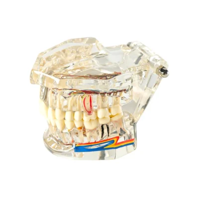

Evodent Signature All in One Model with Nerve Supply Clear, Dental Implant Restoration Model for Patient Education
Original price was: ₹3,500.00 INR.₹1,995.00 INRCurrent price is: ₹1,995.00 INR. -
Sale!

Evodent Four in One Orthodontic Bracket Types Demonstration Model – Metal, Ceramic, Self-Ligating & Lingual Braces
Original price was: ₹10,000.00 INR.₹4,995.00 INRCurrent price is: ₹4,995.00 INR. -
Sale!
")
Evodent Orthodontic Braces vs Aligners Comparison Model for Patient Education (Fixed Appliances vs Clear Aligners)
Original price was: ₹5,000.00 INR.₹2,995.00 INRCurrent price is: ₹2,995.00 INR.
Related products
-
Sale!
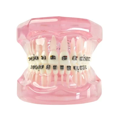

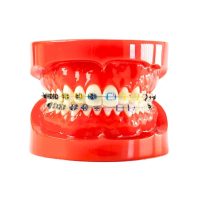
Evodent Orthodontic Patient Education Model with Metal Braces – Pink for Treatment Explanation
Original price was: ₹5,000.00 INR.₹2,495.00 INRCurrent price is: ₹2,495.00 INR. -
Sale!
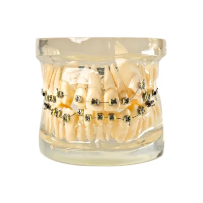

Evodent Signature Dental Malocculation Model with Metal Bracket, Patient Education Model
Original price was: ₹6,000.00 INR.₹3,495.00 INRCurrent price is: ₹3,495.00 INR. -
Sale!
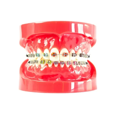

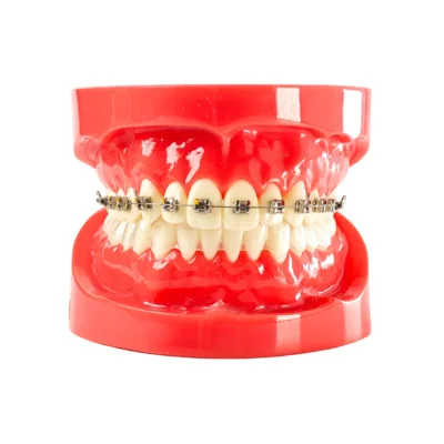
Evodent Orthodontic Model with Metal Braces – Dental Braces Demonstration Model for Orthodontic Treatment Explanation, Red
Original price was: ₹4,000.00 INR.₹1,995.00 INRCurrent price is: ₹1,995.00 INR. -
Sale!
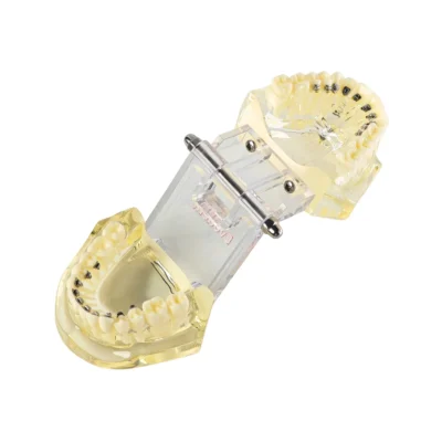

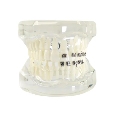
Evodent Lingual Bracket Orthodontic Model ? Lingual Orthodontic Demonstration Model for Patient Education
Original price was: ₹10,000.00 INR.₹3,495.00 INRCurrent price is: ₹3,495.00 INR.